
Ultrasound imaging, or sonography, is a procedure that produces images of the inside of the body using high-frequency sound waves. These images are captured in real-time, and are able to show the structure and movement of blood flow.
What is an ultrasound used for?
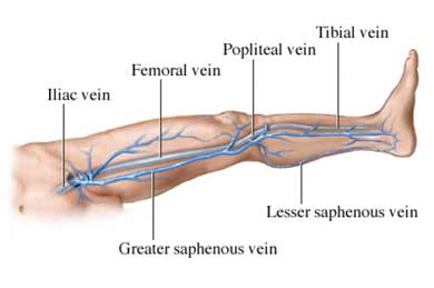
Ultrasound imaging can be used to monitor and diagnose a wide range of conditions. In particular, it is effective at evaluating the location and severity of venous insufficiency. It can also detect deep vein thrombosis and is an important tool during varicose vein procedures.
What happens during the ultrasound?
During the ultrasound, the patient is positioned on an examination table and a clear gel is applied to the area being examined. A transducer is then firmly pressed against the skin and swept back and forth to record the sound waves that are used to obtain the image. The image appears on a computer screen in real time, so that the patient and doctor can view it together.
What are the benefits of an ultrasound?
An ultrasound is a noninvasive, simple procedure that can be used to diagnose many different conditions by producing images of the soft tissues, which often do not show up well on x-rays. There is no ionizing radiation used during this procedure, and is a proven safe diagnostic tool.